Home
/ Arteries Diagram, Arteries Bioninja, Most arteries carry oxygenated blood;
Arteries Diagram, Arteries Bioninja, Most arteries carry oxygenated blood;
Arteries Diagram, Arteries Bioninja, Most arteries carry oxygenated blood;. Learn the differences between an artery and a vein. The aorta branches into a network of smaller arteries that extend throughout the body. 15 diagram of main arteries. The arteries' smaller branches are called arterioles and capillaries. The abdominal aorta bifurcates at the level of the fourth lumbar vertebra to form the two common iliac arteries, each of which further branches into the external and the internal iliac artery.
The heart receives its own supply of blood from the coronary arteries. Inner body parts with their names. Like maps, the various diagrams emphasize different aspects. The right coronary artery courses in the right atrioventricular groove. Is associated with venipuncture, it is done mainly by phlebotomists, nurses, emts and doctors.
Circulatory System Human Anatomy Diagram On Female Body With Arteries And Veins Stock Illustration Download Image Now Istock from media.istockphoto.com These arteries and their branches supply all parts of the heart muscle with blood. Is associated with venipuncture, it is done mainly by phlebotomists, nurses, emts and doctors. Inner body parts with their names. Arteries of the lower limb thigh leg foot the main artery of the lower limb is femoral artery it is a continuation of the external iliac artery terminal branch of the abdominal aorta the arteries and veins of the leg smartdraw arteries and veins of the leg create healthcare diagrams like this example called arteries and veins of the leg in minutes with smartdraw. The vessels make up two closed systems of tubes that begin and end at the heart.one system, the pulmonary vessels, transports blood from the right ventricle to the lungs and back to the left atrium.the other system, the systemic vessels, carries blood from. Arteries carry blood away from the heart in two distinct pathways: Labeled heart diagram showing the heart from anterior. Like maps, the various diagrams emphasize different aspects.
15 diagram of main arteries.
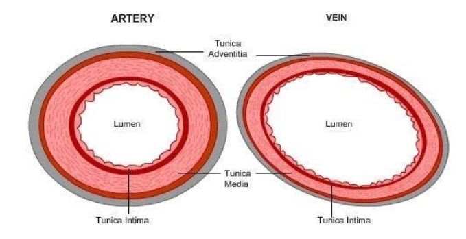
Arteries are blood vessels that carry blood away from the heart. Some are more conceptual, others focus on branching, while still others attempt to preserve a spatial representation. It is returned to the heart in the veins. An artery is an elastic blood vessel that transports blood away from the heart. 5 out of 5 stars. The coronary arteries wrap around the outside of the heart. The narrowed arteries are at higher risk for complete blockage from a sudden. After receiving blood directly from the left ventricle of the heart, the. Inner body parts with their names. Each artery is a muscular tube lined by smooth tissue and has three layers: Most arteries carry oxygenated blood; Arteries of the head and neck diagram art print vintage anatomy art print on tea stained paper dog art dog s wfh office art. Classification & structure of blood vessels.
It is returned to the heart in the veins. Blood is pumped from the heart in the arteries. The right and left subclavian arteries give rise to the thyrocervical trunk. Blood carried by arteries is usually highly oxygenated, having just left the lungs on its way to the body's tissues. The anterior tibial artery forms the arcuate artery and its many branches to supply blood to the top of the foot.
Pin On Study from i.pinimg.com Learn the differences between an artery and a vein. The typical configuration consists of two coronary arteries, a left main coronary artery (lmca) and a right coronary artery (rca), arising from the left posterior and right anterior aortic or coronary sinuses respectively, in the proximal ascending aorta.these are the only two branches of the ascending aorta. These arteries and their branches supply all parts of the heart muscle with blood. More artery diagrams are posted in the following 101 diagramss below. Labeled heart diagram showing the heart from anterior. Anatomy and function of the coronary arteries. Transposition of the great arteries is a serious but rare heart defect present at birth (congenital), in which the two main arteries leaving the heart are reversed (transposed). In this image, you will find external carotid artery, internal carotid artery, vertebral artery, aorta and arch, pulmonary artery, cardiac artery, thoracic aorta, celiac trunk, superior mesenteric artery, renal artery, gonadal artery, inferior mesenteric artery, common iliac artery, external iliac artery.
These vessels are channels that distribute blood to the body.
Coronaryarteriescomplete from faculty.etsu.edu (taken from johnson, weipz and savage lab book). Each artery is a muscular tube lined by smooth tissue and has three layers: After receiving blood directly from the left ventricle of the heart, the. The heart receives its own supply of blood from the coronary arteries. pulmonary artery sling can be treated surgically. Major arteries by definition, an artery is a vessel that conducts blood from the heart to the periphery. Classification & structure of blood vessels. Learn the differences between an artery and a vein. These vessels are channels that distribute blood to the body. An artery (plural arteries) (from greek ἀρτηρία (artēria) 'windpipe, artery') is a blood vessel that takes blood away from the heart to one or more. 15 diagram of main arteries. Arteries and veins of the arm. This is an excellent human heart diagram which uses different colors to show different parts and also labels a number…
Arteries are blood vessels that carry blood away from the heart. The arteries' smaller branches are called arterioles and capillaries. The abdominal aorta bifurcates at the level of the fourth lumbar vertebra to form the two common iliac arteries, each of which further branches into the external and the internal iliac artery. 15 diagram of main arteries. The tunica medica, which is the very muscular middle layer in arteries, is thinner and less muscular in veins.
Draw A Well Labelled Diagram Of Ts Of Artery And Ts Class 11 Biology Cbse from www.vedantu.com Some are more conceptual, others focus on branching, while still others attempt to preserve a spatial representation. After receiving blood directly from the left ventricle of the heart, the. The right coronary artery courses in the right atrioventricular groove. Inner body parts with their names. Smartdraw includes 1000s of professional healthcare and anatomy chart templates that you can modify and make your own. The right and left subclavian arteries give rise to the thyrocervical trunk. 15 diagram of main arteries. An artery is an elastic blood vessel that transports blood away from the heart.
The narrowed arteries are at higher risk for complete blockage from a sudden.
Inner body parts with their names. The tunica medica, which is the very muscular middle layer in arteries, is thinner and less muscular in veins. Bodytomy provides a labeled iliac artery diagram to help you understand the anatomy and function of the common iliac. This is a congenital defect in which the left pulmonary artery branches off the right pulmonary artery, rather than directly from the pulmonary trunk. Transposition of the great arteries is a serious but rare heart defect present at birth (congenital), in which the two main arteries leaving the heart are reversed (transposed). Each of these arteries delivers blood to the leg and continues into the foot, with the posterior tibial and fibular arteries forming the plantar arteries and plantar arch that supply blood to the bottom of the foot and toes. Labeled heart diagram showing the heart from anterior. Two major coronary arteries branch off from the aorta near the point where the aorta and the left ventricle meet. Blood vessels are the channels or conduits through which blood is distributed to body tissues. The typical configuration consists of two coronary arteries, a left main coronary artery (lmca) and a right coronary artery (rca), arising from the left posterior and right anterior aortic or coronary sinuses respectively, in the proximal ascending aorta.these are the only two branches of the ascending aorta. The right coronary artery courses in the right atrioventricular groove. The coronary arteries wrap around the outside of the heart. It is returned to the heart in the veins.